


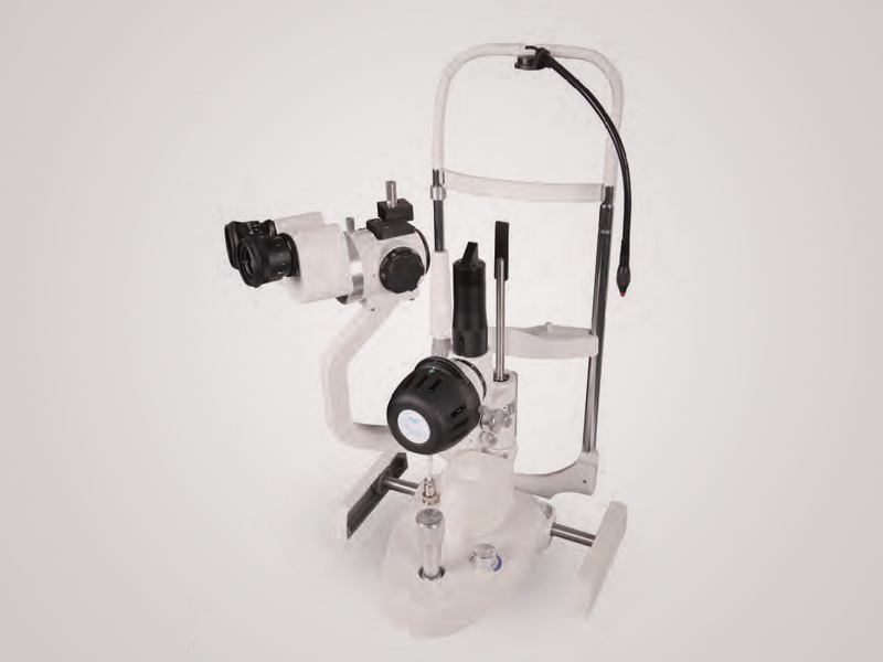
S260/S260S SLIT LAMP MICROSCOPE
- Classic galilean type microscope
- Magnification Ratio:
- 6x, 10x, 16x, 25x, 40x (S260)
- 10x, 16x, 25x (S260S)
- Max. Aperture diameter : O 14mm
- Powered by led light source
- Convergence angle of eyepieces : 13o
- Excellent Scalability, Compatible with Applanation Tonometer, Digital Camera, CCD, teaching tube, etc
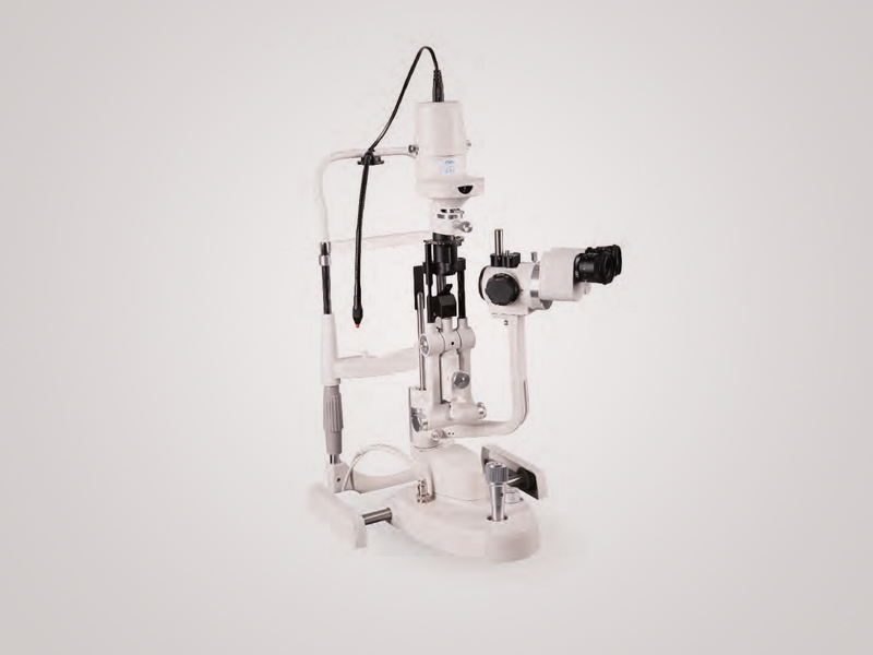
S 360 / S360S SLIT LAMP MICROSCOPE
- Classic galilean type microscope
- Total Magnification Ratio:
- 6x, 10x, 16x, 25x, 40x (S360)
- 10x, 16x, 25x (S360S)
- Max. Aperture diameter : O 14mm
- Powered by led light source
- Convergence angle of eyepieces : 13o
- Excellent Scalability, Compatible with Applanation Tonometer, Digital Camera, CCD, teaching tube, etc.
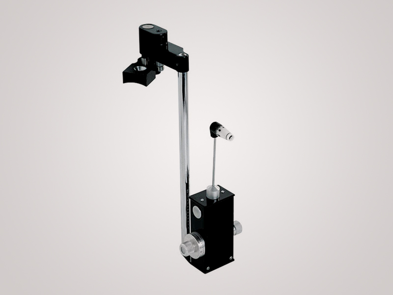
Applanation Tonometer T 170
- Gold Standard
- Accurate Measurement
- Excellent Mechanics
- Premium Optics
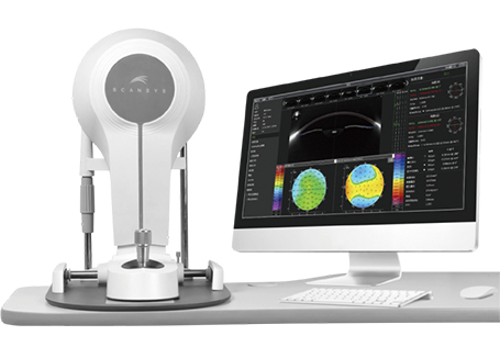
Scansys – Anterior Segment Analyser
- Basic model
- 28 Cornea Tomography Images
- Anterior Chamber Depth
- Total Cornea Power
- Refractive 4 Maps
- Aberration Analysis
- Refractive Power Distribution
- 2D/3D Data Collection
- True Net Refractive Power
- Refractive Power (Front)
- AI Keratoconus Analysis
- Pachymetric Distribtion
- Lens Fitting
- Keratometric Power Deviation
- Cornea Thickness Maps
- Cornea Curvature / Elevation Maps
- Chamber Angle Analysis
- Lens Density Analysis
- Form Factor
Professional model
- 60 Cornea Tomography Images
- IOL Calculation
- IOL Optimization
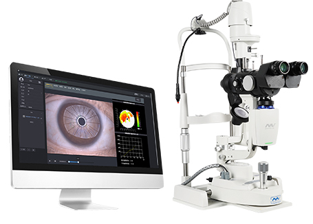
S 390L Slit Lamp (Firefly WDR) with Dry Eye Diagnostic System
- Simple Design + Simple Operation
- Built in Yellow Filter
- The Meibomian Glands Examination
- Convenient Patient Management
- Functional Image Analysis
- Orthokeratology Lens Fitting Assistance
- Customized Auto Exposure Value Setting
- The Most Effective Tools for Dry Eye
- Meibomian Gland Observation
- Tear Film Break-up Time
- Red Eye Analysis and Keratopathy Exposure
- Tear Meniscus Height
- Lipid Layer Thickness
- Eyelid Margin
- AI Analysis of Conjunctival Hyperemia
- Cornea Sodium Fluorescein Staining
- Dry Eye Comprehensive Evaluation Report
- Smart Patient Management System
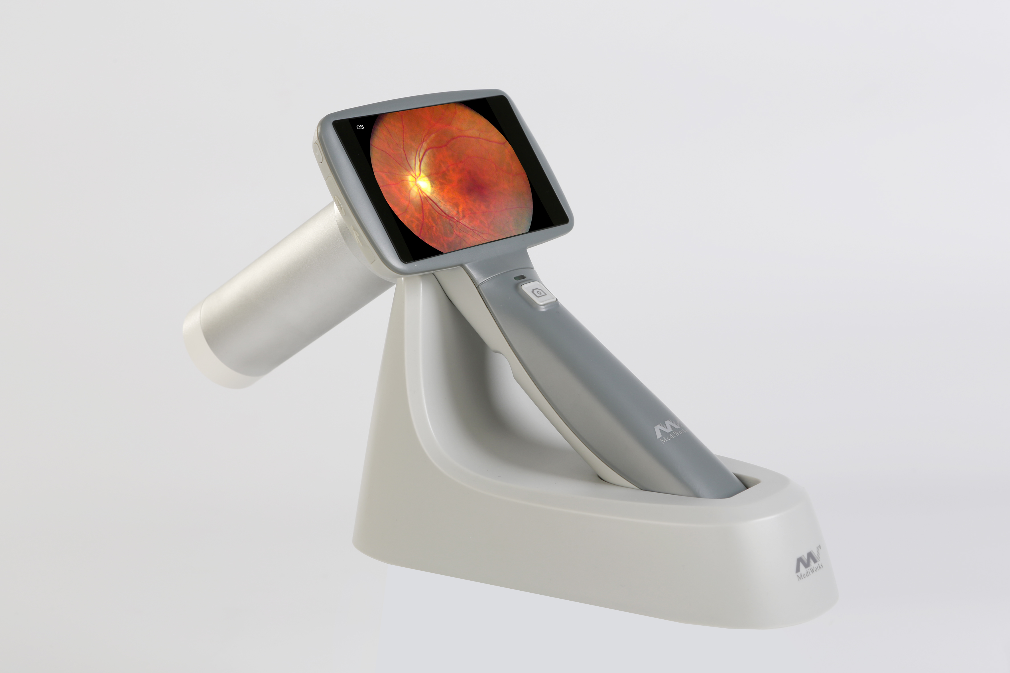
FC 161 Hand Held Fundus Camera
- Auto Split Focusing Technology Millisecond Focusing Speed
- High Resolution
- Non Mydriatic
- 9 Fixation Targets
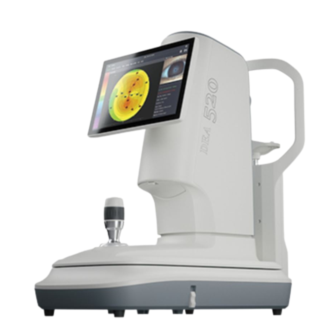
DEA 520 – 2 IN 1 Ocular Diagnostic Master – Corneal Topographer
- 1 Ring
- 3 illuminations
- 9 functions
Placido Ring
- Thousands of points measurements - ensure more data available and accurate analysis
- Smaller cone design – bigger projection area
- 3 illuminations – white illumination, infrared illumination, cobalt blue illumination
9 functions
Dry Eye Diagnosis
- Non Invasive Tear Film Break Up Time
- Cornea Sodium Fluorescein Staining
- Non – Invasive Tear Meniscus Height
- Eyelid Margin
- Meibomian Glands Function Evaluation
- Conjunctival Redness Analysis
- Lipid Layer Thickness
Topography
Topography Analysis
